
Age and Decreased Ovarian Reserve
Most women will experience a decline in fecundability as they age that
is physiologic rather than pathologic. This decline begins in the early
30s and accelerates during the late 30s and early 40s, reflecting
declines in oocyte quantity and quality. Among populations that do
not practice contraception, fertility peaks at age 20, declines somewhat at age 32, steeply declines after the age of 37, and is rare after age 45. An association between the age of the woman and reduced fertility is well documented. Disorders of ovulation account for about 20% to 40% of all cases of female infertility.
Although we expect the ovary to age in a certain way, there are times when it doesn't behave as predicted. That's why screening for ovarian reserve is a fundamental part of the initial evaluation for infertility patients of any age.
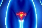
What is Ovarian Reserve
The term "ovarian reserve" refers to a woman's current supply of eggs, and is closely associated with reproductive potential. In general, the greater the number of remaining eggs, the better the chance for conception. Conversely, low ovarian reserve greatly diminishes a patient's chances for conception.
Since a woman's chronological age is the single most important factor in predicting a couple's reproductive potential, age has often guided infertility treatment choices. However, age alone doesn't
tell the whole story.
Methods of Assessing Ovarian Reserve
The methods for assessing ovarian reserve are classified into two groups: passive testing and dynamic testing. The goal of both approaches is to provide information regarding oocyte (egg) quality and quantity. Methods for assessing fertility potential include the measurement of FSH, LH, estradiol, inhibin-B, and Antimullerian hormone (AMH).
Early follicular-phase FSH levels play an important role in pregnancy outcomes and day 3 FSH could be very useful in predicting response to ovulation induction. As a woman ages, FSH becomes
elevated in an attempt to force the aging ovary to respond. A rise in early follicular-phase FSH is also accompanied by a decline in oocyte quality, and some investigators have linked such FSH
elevations to fetal abnormalities. FSH values emerged as superior to maternal age as a method for determining reproductive outcome.
However, a single measurement of day 3 FSH may not represent actual ovarian reserve. LH measurement may also have predictive value for ovarian reserve. There may be a place for combined FSH+LH testing to estimate ovarian reserve, as some investigators have suggested an increased FSH:LH ratio may predict an elevation in FSH alone.
Basal day 3 FSH level is often combined with estradiol (E2) testing. Estradiol levels on day 3 of the menstrual cycle reflect follicular growth rather than the number of antral follicles. Elevations in FSH and decreases in inhibin B that accompany aging results in advanced follicular growth at the end of the preceding luteal phase.
Serum inhibin B is secreted by ovarian granulosa cells starting at the preantral follicle stage and therefore reflects the size of the growing follicular cohort. Reduced inhibin B levels are seen with aging even in normal fertile women. Inhibin B alone has poor predictive value for ovarian response but improves the predictive value when added to the CCCT.
Antimullerian hormone (AMH) is produced by the granulosa cells of preantral and small antral follicles. The serum evel of AMH in women with normal cycles declines with age and becomes undetectable
after menopause. AMH appears to be a good predictor of both excessive (>3.5 ng/mL) and poor. Unlike other serum markers, AMH can be measured at any time in the menstrual cycle.
Dynamic Ovarian Reserve Testing
In contrast to the static measurements of ovarian reserve mentioned, the clomiphene citrate challenge test (CCCT) is a dynamic approach. Its purpose is to stimulate the ovary to initiate egg
production in response to a fertility drug called clomiphene (Clomid or Serophene). In theory, the CCCT was designed to detect low ovarian reserve that would not be discovered by a single FSH and/or
E2 measurements.
The CCCT is based on the assumption that adequate ovarian reserve is associated with a healthy group of developing follicles. This healthy group of follicles should be capable of producing enough
inhibin and E2 to suppress FSH production and resist the effects of clomiphene.
Clomiphene works by shutting down the estrogen receptors on the hypothalamus and tricking the hypothalamus into thinking the patient doesn't have enough estrogen. In response, the hypothalamus works
harder to induce the pituitary gland to produce more FSH and LH. This, in turn, initiates follicular growth.
Antral Follicle Count: Using transvaginal ultrasound in the early follicular phase, all ovarian follicles 2 to 10 mm are counted and the total for both ovaries is called the basal antral follicle count (AFC). The AFC correlates well with chronologic age in normal fertile women and appears to reflect what remains of the primordial follicular pool. Decreases in AFC with age are gradual rather than sudden. A total AFC less than 4 is predictive of poor response.
Contact Us
Chalmers Medical Building
328 Hwy 7 East Suite 201,
Richmond Hill ON L4B 3P7
Tel: 416 399-3888
E-mail: info@aahclinic.com

Conditions Treated


Effective Herbal Therapy for Inflammatory Bowel Disease
NO Surgery!
Carolyn XU Treats Ulcer Colitis Successfully
Considering Therapy?
Resource Links


References


Donation
Great things happen when you put your heart into it.


Click Here to Send us your request
Tips
Questions about your first appointment or if your insurance will cover the cost? Find more information below.